|
|
|
|
|
|
|
|
|
|
|
|
|
Take the Test |
It is important to remember that there is a wide range of normal variability in the 12 lead ECG. The following "normal" ECG characteristics, therefore, are not absolute. It takes considerable ECG reading experience to discover all the normal variants. Only by following a structured "Met hod of ECG Interpretation" (Lesson II) and correlating the various ECG findings with the particular patient's clinical status will the ECG become a valuable clinical tool.
Topics for Study:
How to calculate the heart rate on ECG paper
Because ECG paper moves at a standardized 25mm/sec, the vertical lines can be used to measure time. There is a 0.20 sec between 2 of the large lines. Therefore, if you count the number of heart beats (QRS complexes) in between 30 large boxes (6 seconds) and multiply by 10, you have beats per minute. Conveniently, ECG paper usually has special markings every 3 seconds so you don't have to count 30 large boxes.
There is, however, an easier and quicker way to estimate the heart rate. As seen in the diagram below, when QRS complexes are 1 box apart the rate is 300 bpm. 2 boxes apart...150 bpm, etc. So if you memorize these simple numbers you can estimate the heart rate at a glance!
QT < 0.38 @ 80 bpm
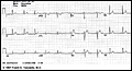
(Normal ECG is shown below - Compare its waveforms to the descriptions below)
Click to view
![]() QRS Complex
QRS Complex
The QRS represents the
simultaneous activation of the right and left ventricles, although most
of the QRS waveform is derived from the larger left ventricular musculature.
![]() QRS duration < 0.10 sec
QRS duration < 0.10 sec ![]() QRS amplitude is quite variable from lead to lead and from
person to person. Two determinates of QRS voltages are:
QRS amplitude is quite variable from lead to lead and from
person to person. Two determinates of QRS voltages are:
Click to view
Click to view
Click to view
Click to view
![]() The normal U Wave: (the most neglected of the ECG waveforms)
The normal U Wave: (the most neglected of the ECG waveforms)
![]() U wave amplitude is usually < 1/3 T wave amplitude in same
lead
U wave amplitude is usually < 1/3 T wave amplitude in same
lead ![]() U wave direction is the same as T wave direction in that lead
U wave direction is the same as T wave direction in that lead
![]() U waves are more prominent at slow heart rates and usually best
seen in the right precordial leads.
U waves are more prominent at slow heart rates and usually best
seen in the right precordial leads. ![]() Origin of the U wave is thought to be related to
afterdepolarizations which interrupt or follow repolarization.
Origin of the U wave is thought to be related to
afterdepolarizations which interrupt or follow repolarization.

)
)
)
)
)